Journal of Current Surgery, ISSN 1927-1298 print, 1927-1301 online, Open Access
Article copyright, the authors; Journal compilation copyright, J Curr Surg and Elmer Press Inc
Journal website http://www.currentsurgery.org

Case Report

Volume 2, Number 2, April 2012, pages 62-65


Infected Bilateral Chylothorax in a Problematic Case

Figures

Figure 1.
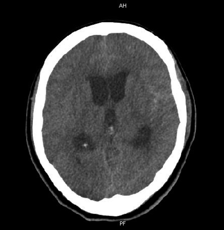
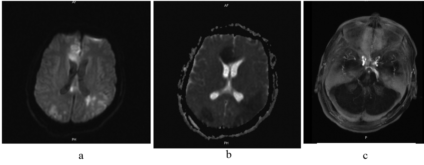
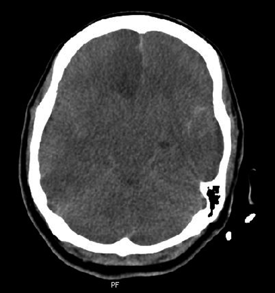
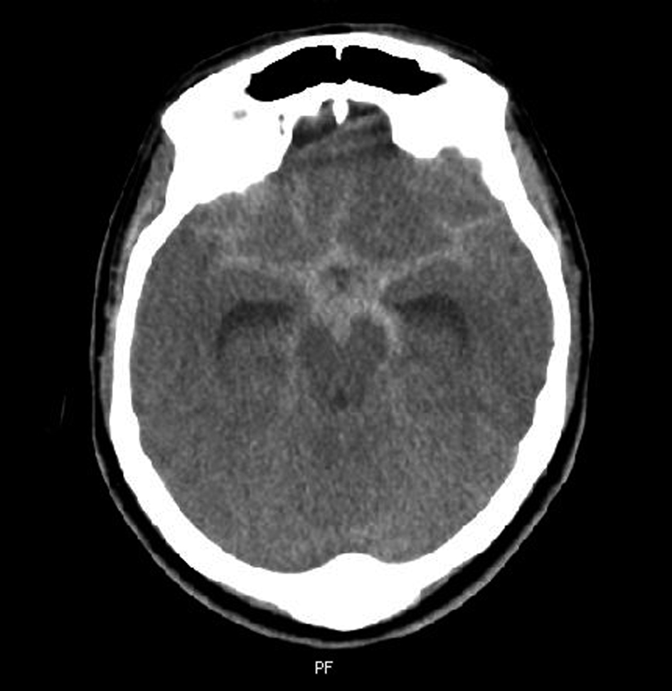
Figure 1. Cranial CT scan discloses subarachnoid haemorrhage.
Figure 2.
Figure 2. MR-angiography reveales a berry aneurysm at the tip of the basilar artery.
Figure 3.
Figure 3. CT scan reveales obstructive hydrocephalus.
Figure 4.
Figure 4. Chest X-ray shows bilateral massive pleural effusions.
Figure 5.
Figure 5. Chest CT discloses mediastinal enlargement.
Figure 6.
Figure 6. (6a and b) Diffusion and perfussion weighted cranial MR show bilateral posterior parietal, and right medial frontal hemispheric ischemia; and (6c) MR-angiography discloses vasospasm at both right anterior cerebral artery and bilateral middle cerebral arteries.
Figure 7.
Figure 7. Cranial CT shows bilateral cerebral oedema with subfalcine herniation towards to left side.