Figures
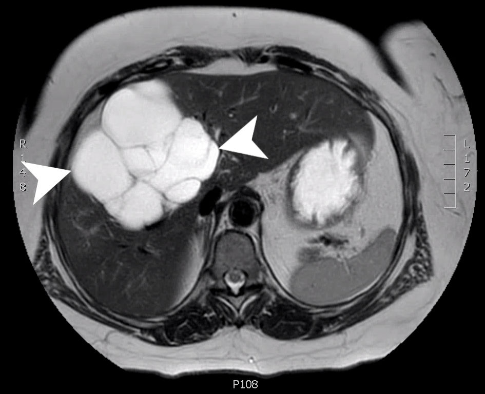
Figure 1. An axial slice from a T2 weighted MRI with gadolinium contrast showing a complex, multilocular cystic mass (arrow heads) measuring 10.4 × 10.2 × 14.1 cm involving segments 8, 5 and 4B. MRI: magnetic resonance imaging.
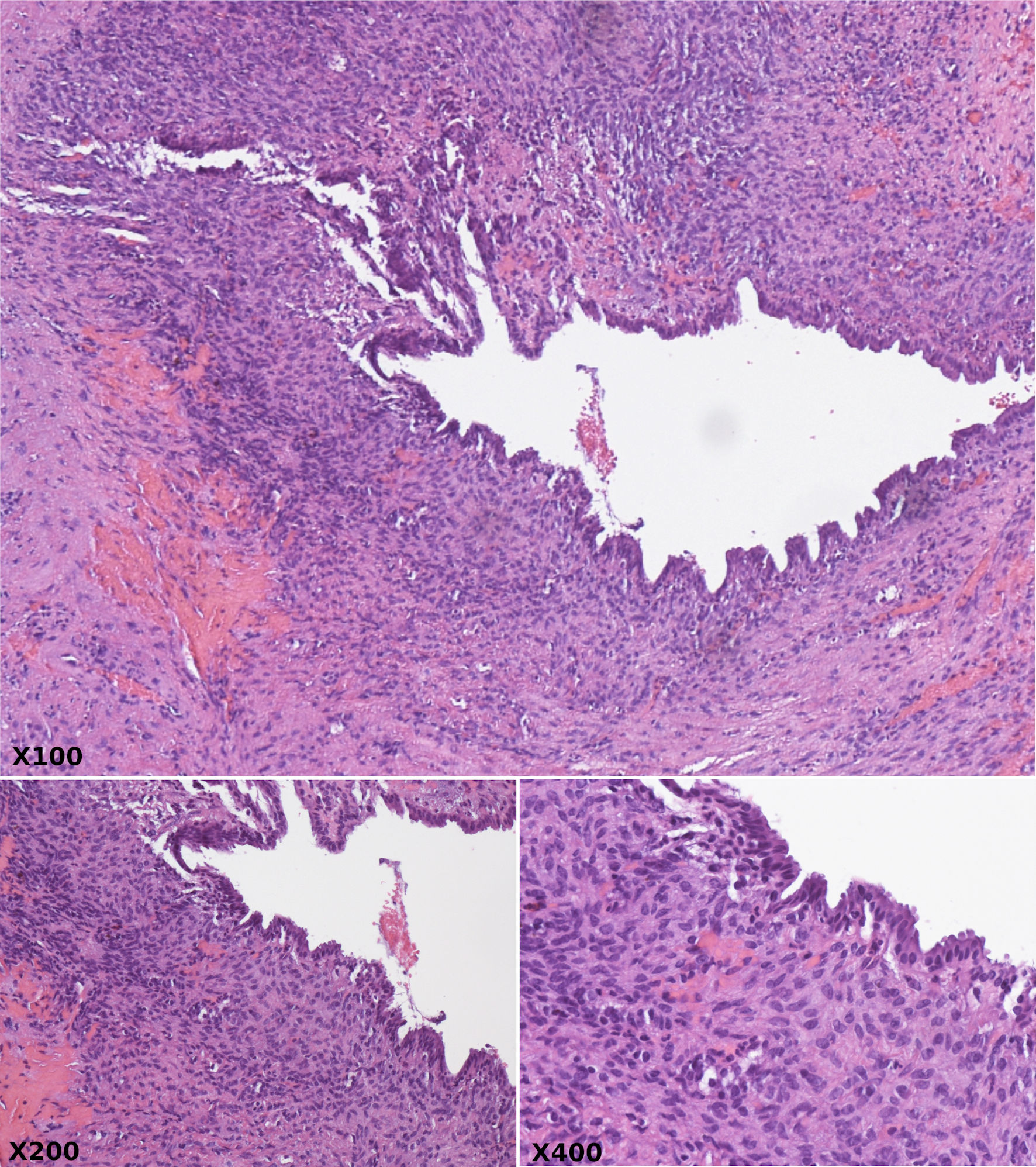
Figure 2. Hematoxylin and eosin stain at × 100, × 200, and × 400 magnification revealing cystically dilated ducts lined by bland biliary columnar epithelium and surrounded by compact ovarian type spindle cell stroma.
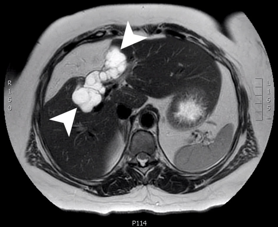
Figure 3. An axial slice from a T2 weighted MRI scan with Eovist (gadoxetate disodium) contrast. A multiloculated cystic mass (arrow heads) is seen involving the right hepatic lobe (segments 4 and 5) measuring 8.0 × 3.2 × 3.2 cm. MRI: magnetic resonance imaging.
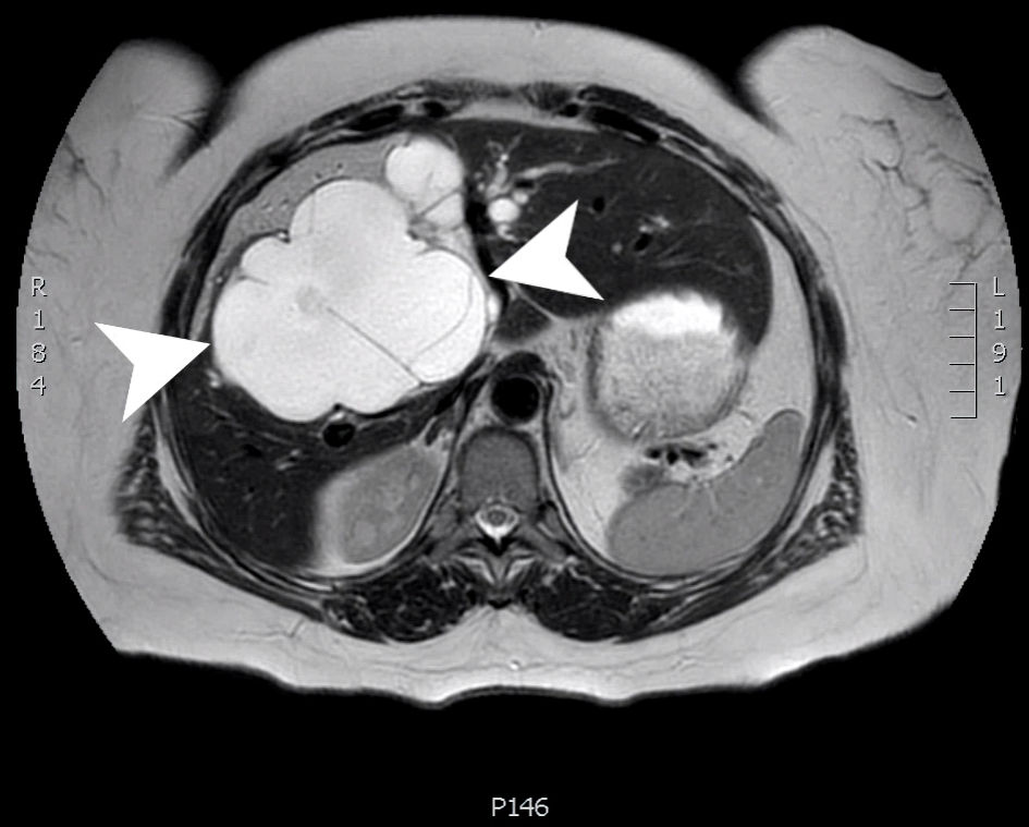
Figure 4. An axial slice from a T2 weighted MRI scan with Eovist contrast. A large, multiloculated cystic structure (arrow heads) within the central liver (segments 4, 5 and 8) measuring 12.5 × 8.9 × 2.1 cm. MRI: magnetic resonance imaging.
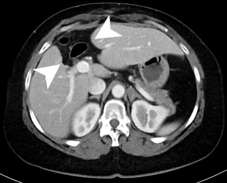
Figure 5. An axial slice from the venous phase of a CT scan with contrast. The scan shows chronic postoperative changes of a prior central hepatectomy within the inferior aspects of segments 4B and 8 (arrow heads) with evidence of compensatory hypertrophy of both the right and left hemi-liver. CT: computed tomography.