Figures
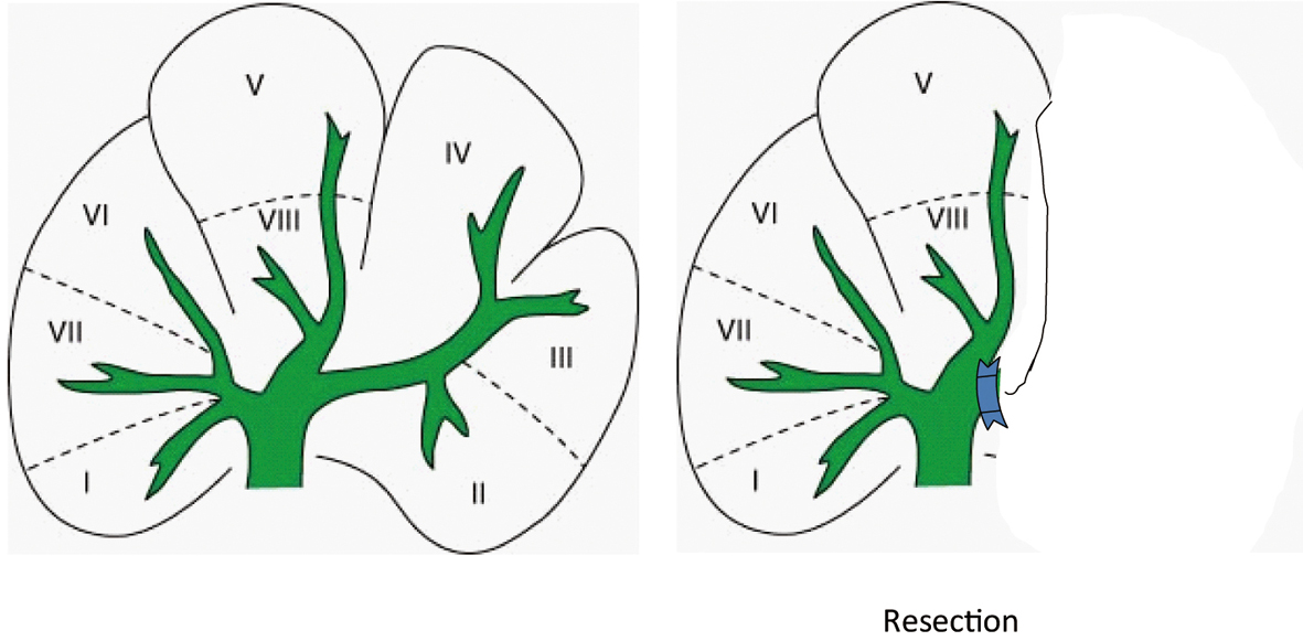
Figure 1. The left portal vein and artery was ligated, and a left hemi-hepatectomy was performed (approximately 40%).
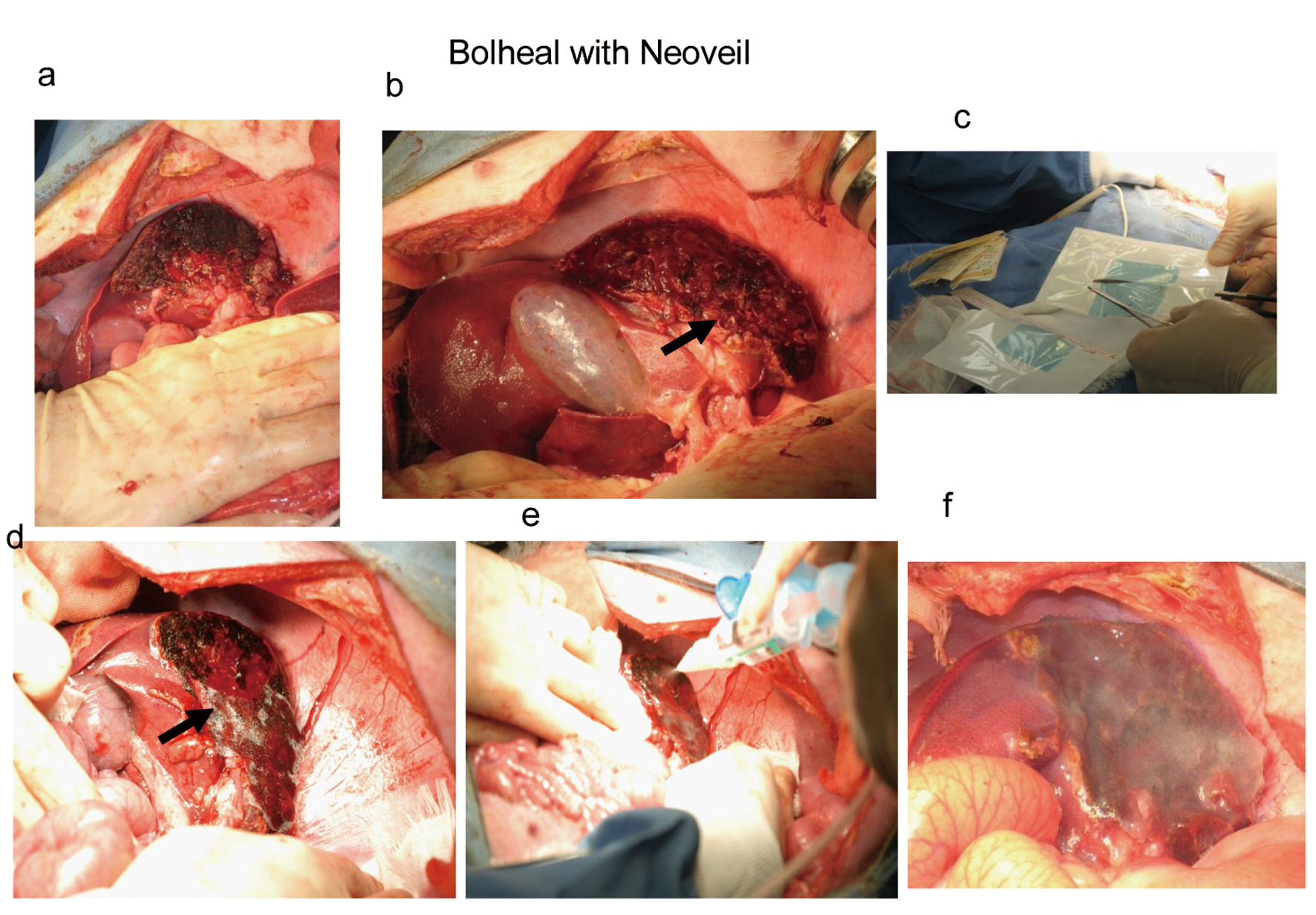
Figure 2. After controlled bleeding and bile leakage (a), solution A was applying to the cut surface (b), then 1 cm2 piece of Neoveil (c) were attached covering the surface (arrow in d), and solution A and B were applied (e and f).
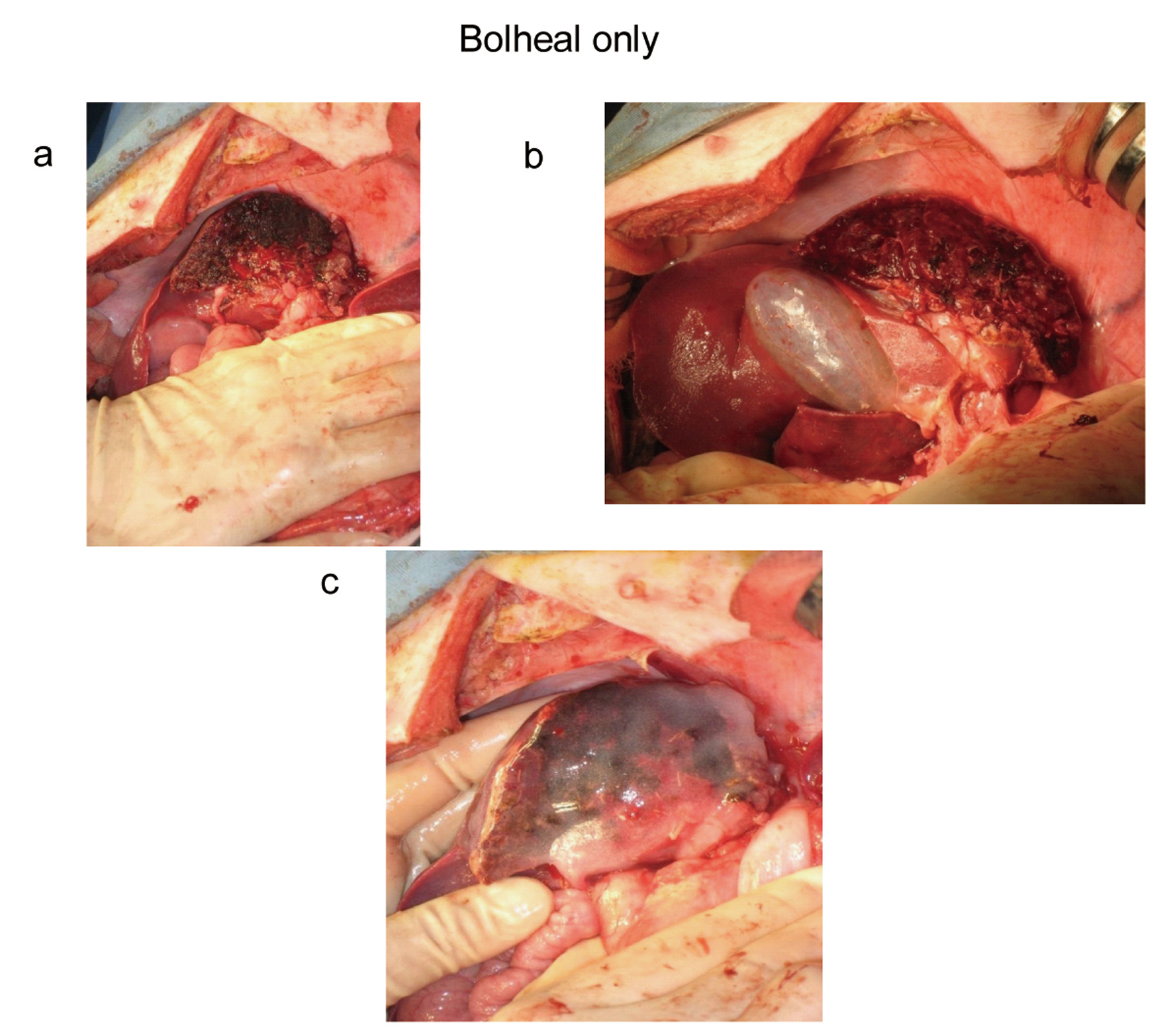
Figure 3. After liver resection, we controlled bleeding and bile leakage (a). Then, 1 mL solution A was applied to the cut surface (b), and then followed by solution A and B applied to the covered surface (c).
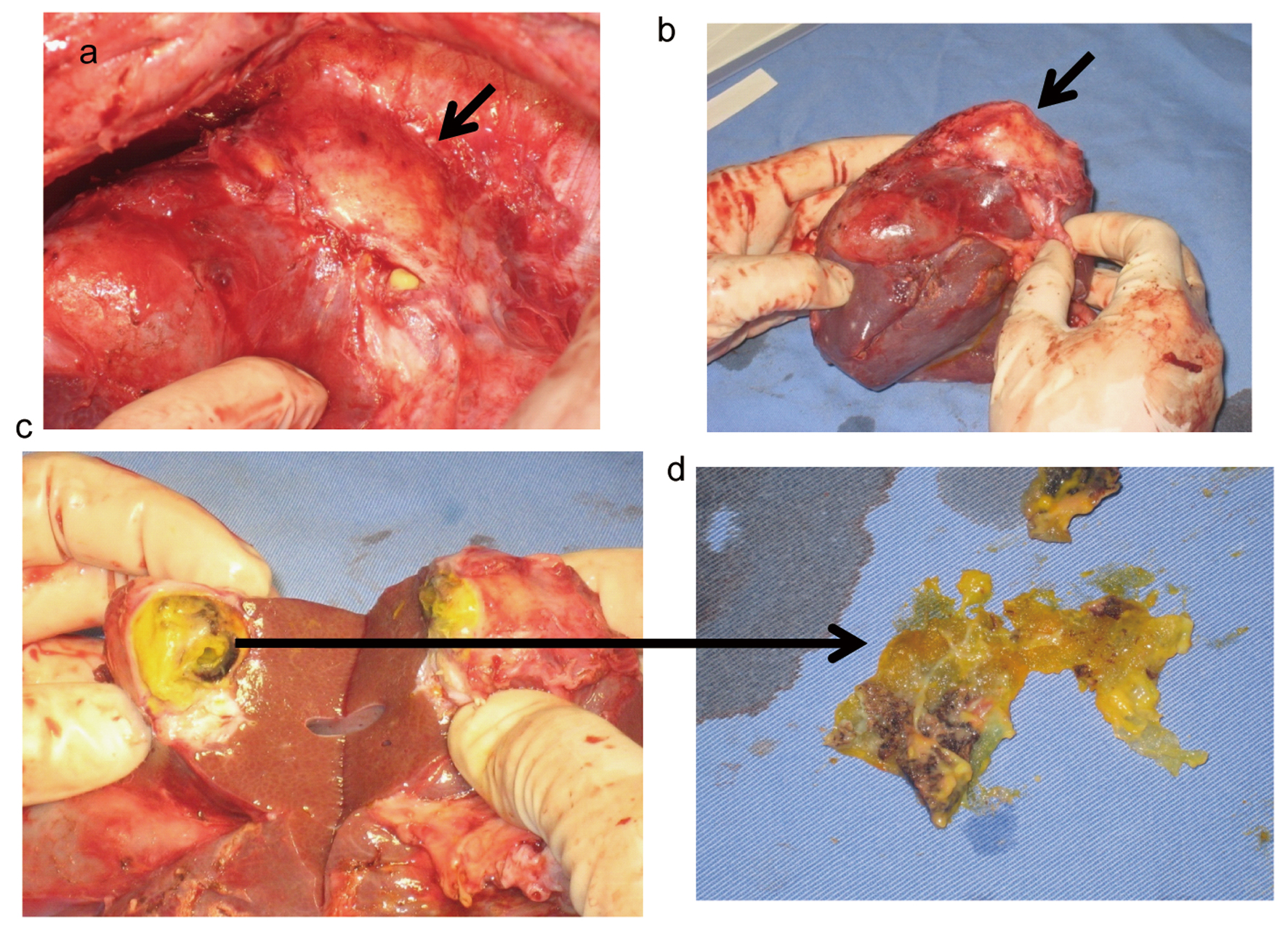
Figure 4. Fibrotic capsulated cavity revealed at the cut surface (a, b: arrows). Inside of this cavity had necrotic liver tissues, Bolheal, and peace of Neoveil with bile juice (c, d).
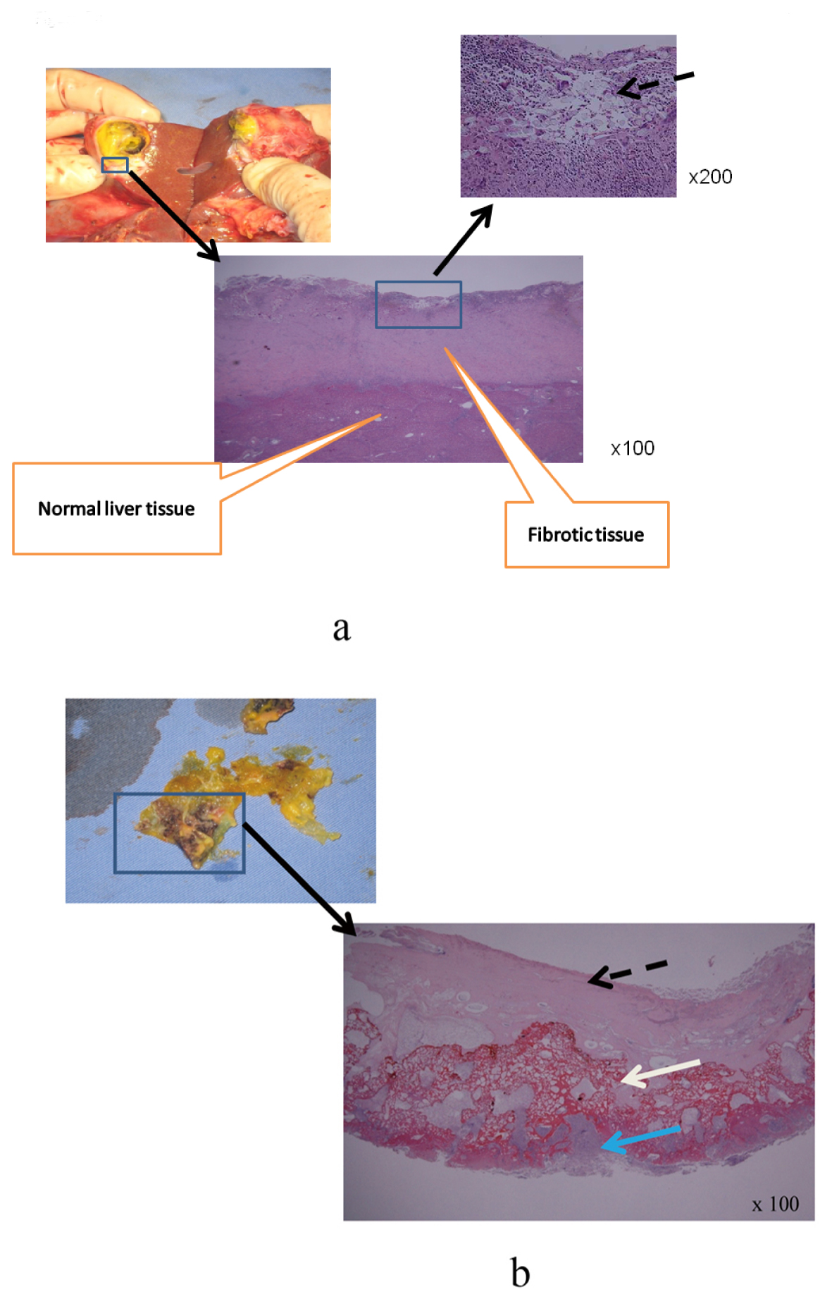
Figure 5. a: The fibrotic tissue and normal liver tissue were completely separate; Bolheal and Neoveil were remained on the fibrotic tissue (dot arrow); b: Neoveil, Bolheal and carbonizing liver tissue were not adapted, Bolheal; dot arrow, Neoveil; white arrow, and necrotic liver tissue (blue arrow), respectively.
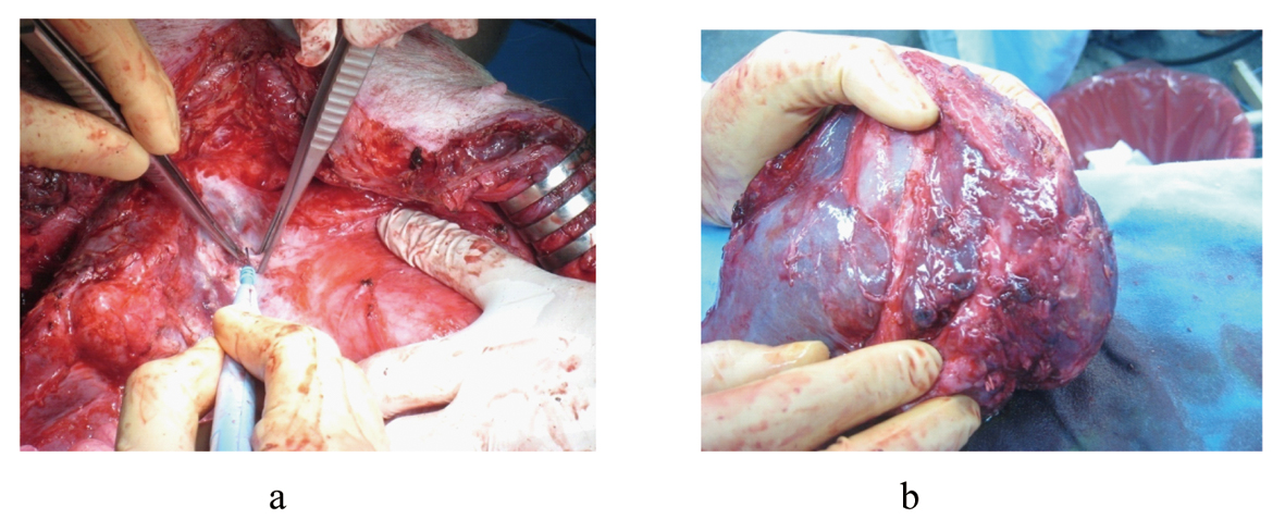
Figure 6. Mild adhesion revealed at the cut surface (a) and specimen did not had any kind of biloma or abscess.
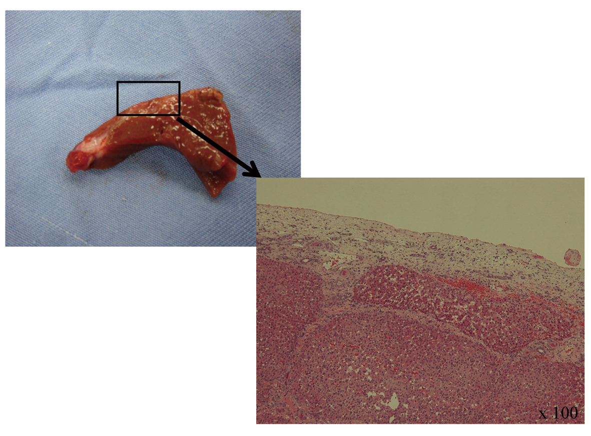
Figure 7. Histology, Bolheal was integrated with normal liver tissues, Bolheal did not find anywhere at the cut surface, fibrotic tissue was covered normal liver tissue.